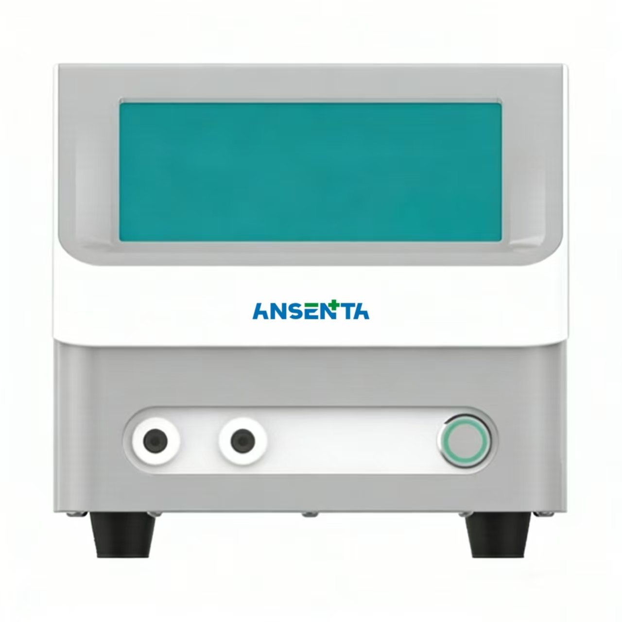
Product Description
The GP-3000 Gene Electroporator consists of the main unit, gene electroporation cup, and dedicated connecting cables. It primarily uses electroporation to introduce DNA into competent cells, plant and animal cells, and yeast cells. Compared to other methods, the gene electropora- tor offers high repeatability, high efficiency, ease of operation, and precise control. Additionally, electroporation has no genotoxicity, making it an essential technique in molecular biology.
Working Principle
Cell electroporation (also called cellular electroporation) is an important method for introducing exogenous macromolecules such as DNA, RNA, siRNA, proteins, and small molecules into cells.
Under the influence of a strong, momentary electric field, the cell membrane in the solution becomes permeable, allowing charged exogenous substances to enter the cell membrane in a manner similar to electrophoresis. Due to the high resistance of the cell membrane’s phospholipid bilayer, the voltage produced by the external electric field is borne by the cell membrane, with minimal voltage distributed to the cytoplasm, result- ing in negligible intracellular current and low cytotoxicity during electroporation. After DNA or other substances enter the cell membrane, they remain near the membrane, with subsequent diffusion into the nucleus or other parts facilitated by the cell’s mechanisms.
Since electroporation is a physical method, the molecular properties of the cell surface have little effect on the process. Compared to chemical transfection methods or viral vector transfection, electroporation can be applied to all cell types and is easy to quantify.
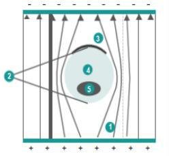
Cell Electroporation Electric Field Diagram
1 The cell membrane behaves like an insulator, causing current distortion in the electroporation fluid near the cell.
2 In a series circuit, the larger the resistance, the higher the voltage; most of the voltage is borne by the cell membrane at both ends.
3 Only one end of the cell is effectively electroporated.
4 The voltage distributed to the cytoplasm is very small; DNA stops at the membrane after electro- poration, and its subsequent diffusion is driven by the cell’s natural mechanisms.
5 The voltage at both ends of the nucleus is extremely small, and this small voltage is borne by the nuclear membrane, leaving the interior of the nucleus completely voltage-free, which eliminates genotoxicity during electroporation.
Product Features
High Efficiency Short transformation time, high conversion rate, excellent repeatability.
Intelligent Storage Experimental parameters can be stored and easily retrieved by users
Precise Control Pulse discharge is controlled by a microprocessor.
Aesthetic Design Integrated design, intuitive display, and easy operation.

Application Areas

· Electroporation of bacteria, yeast, and other microorganisms.
· Transfection of mammalian cells, plant tissues, and protoplasts.
· Cell hybridization and gene fusion.
· Introduction of marker genes for labeling and indicating purposes.
· Introduction of drugs, proteins, antibodies, and other molecules for studying cellular structure and function.
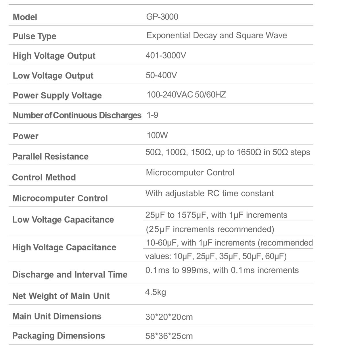
Technical Parameters
Conversion Rates for Different Strains
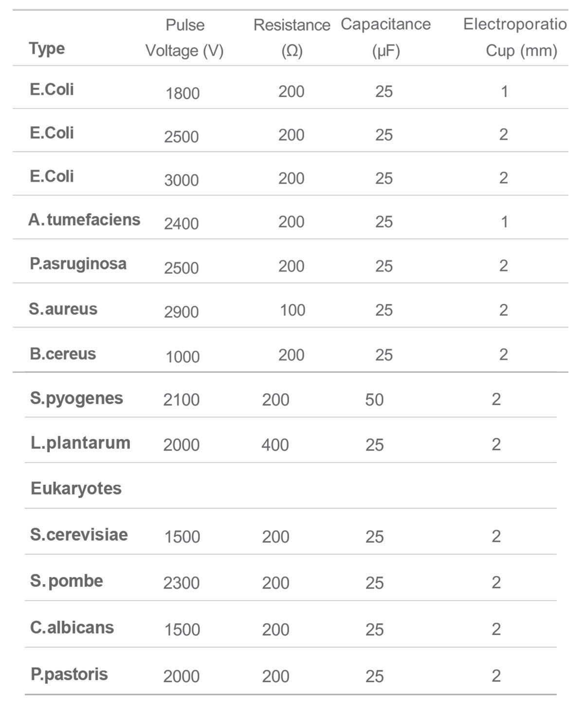
Note: Due to variations in experimental conditions across different labs,the above parameters are for reference only.
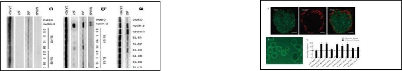
Example Experiments
1、LIJ, ZHANG S, GAO L, et al. A cell-based high-throughput assay for the screening of small-molecule inhibitors of p53-MDM2 interaction [J]. Journal of biomolecular screening, 2011.
2、ZHANG SY, LIJ, XIE X. Discovery and characterization of novel small molecule agonists of G protein-coupled receptor 119 [J]. Acta pharmacologica Sinica, 2014.